Overview –
1. SCALP layer’s
2. Meninges
3. Extra cranial or SCALP haemorrhage
4. Intra cranial hemorrhage
5. Cause
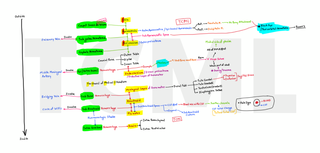
Layers from outside to inside –
1. Skin
2. Connective tissue
3. Aponeurosis or epicranial aponeurosis or galea aponeurotica
4. Loose connective tissue / Sub aponeurotic space
5. Pericardium / Outer periosteum
6. Outer table
7. Diploe
8. Inner table
9. Endocranium / Inner periosteum or Periosteal layer / Endosteal layer of dura mater
10. Meningeal layer of dura mater
11. Arachnid
12. Piamater
13. Brain
(for more detail see diagram)
NOTE – In these 13 points
1. SCALP layer’s from 1 to 5
2. Skull bone layer from 6 to 8
3. Meninges from 9 to 12

Clinical –
Extra cranial hemorrhage from 1 to 3
Intra cranial hemorrhage from 4 to 7
(See diagram)
1. Caput succedaneum –
● It is a localized edema that is present on scalp region of newborn.
● Caput succedaneum need no treatment because it disappear spontaneous.
2. Subgaleal hematoma –
● As the clear by name it is present below the galea aponeurotica.
NOTE – Black Eye or Periorbital hematoma
3. Cephalo hematoma –
● Collection of blood between outer periosteum and outer table of skull bone.
● Cephalo hematoma is limited by suture line so it is never present over the suture line.
4. Epidural hemorrhage –
● In CT scan, this hematoma looks biconcave shape.
● Lucid interval present
NOTE – Pterion
● It is an example of epidural hemorrhage.
● It is area of skull and four skull bones frontal, parietal, temporal, and sphenoid meet here and form H-shape suture.
● Anterior division of middle meningeal artery (MMA) is present on the medial side of the pterion. That’s why due to the fracture of pterion, the anterior division of MMA is ruptured and causes epidural hemorrhage.
For more detail about pterion click here
5. Subdural hemorrhage –
● It is present between dura mater (meningeal layer) and arachnoid.
● In CT scan, this hematoma looks crescent shape.

6. Sub arachnoid hemorrhage (SAH) –
● SAH is present in subarachnoid space.
● Lumbar puncture – Because in the case of SAH, blood mix with CSF. That’s why we examine CSF through lumbar puncture.
● Xanthochromia – Blood mix in the CSF and CSF color was change (Yellow color CSF)
● Thunderclap headache (acute single episode of headache) present
For more detail about Subarachnoid Haemorrhage (SAH) click here
7. Intra cerebral hemorrhage –
● In this case hemorrhage present inside the brain tissue and brain ventricle.
Causes of cranial hemorrhage –
1. Birth injury – Due to external pressure on newborn head by birth canal during vaginal delivery. (Obestetrics)
2. Vacuum assited delivery or Ventouse (Obestetrics)
3. Forceps delivery (Obestetrics)
4. Skull bone fracture
5. Trauma
6. Hypertension
7. Stroke
8. Head injury, Brain injury, Skull injury
9. Neoplasm
10. Drug abuse
11. Angiopathy (Disease of blood vessels) –
● Cerebral Amyloid Angiopathy (CAA) – Amyloid beta peptide (A beta)
● Diabetic angiopathy
12. Cerebral Aneurysm –
● Due to weak blood vessel wall, abnormal localized dilated present on the vessel wall.
● Bleeding occurs due to rupture of aneurysm.
NOTE – Saccular aneurysm most common type.
13. Venous thrombosis
🔴 Now all TCML hand written notes and charts are available on TCML Mobile App (FREE for all users).
To download TCML Mobile App click on below pic.

Brain blood supply – Vertebral Artery, basilar artery, internal carotid artery, and circle of willis (COW)