Overview-
1. Boundaries- Anterior, posterior, superior, inferior, lateral, and medial.
2. Communication- Infratemporal fossa.
3. Content- Muscle, artery, and nerve.
● It is a fan shape, shallow fossa.
● Two in number and present on anterolateral side of the skull.
● Temporalis muscle (Fan shape muscle) are attached on it.
1. Boundaries-
The boundaries of temporal fossa are:
A. Anterior-
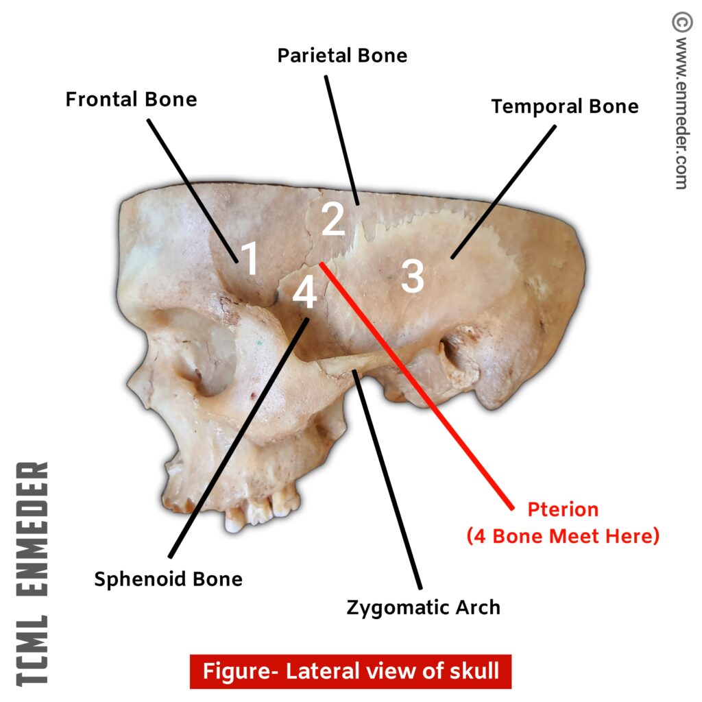
It is formed by frontal and zygomatic bone. (See Fig. Lateral view of skull)
B. Posterior-
It is formed by Inferior temporal line and supra mastoid crest.
C. Superior-
It is formed by Superior temporal line.
D. Inferior-
Inferiorly it is communicate with infra temporal fossa. (See communication of temporal fossa)
E. Lateral or Roof-
Temporal fascia, zygomatic arch form lateral boundary of temporal fossa.
F. Medial or Floor-
It is formed by the part of four skull bones: Frontal, parietal, temporal, and sphenoid bone (Greater wing). (Fig. Lateral view of skull)
Pterion– See point 4 (Clinical)
2. Communication-
It is communicate below with infra temporal fossa through a space deep to the zygomatic arch. (See inferior boundaries of temporal fossa)

3. Content-
A. Muscle-
It contain the temporalis muscle (Muscle of mastication)
B. Artery-
● Middle meningeal Artery (Branch of superficial temporal artery)
● Deep temporal artery (This is the branch of 2nd part of maxillary artery)
● Zygomatico temporal artery (arise from Zygomatic branch of Lacrimal artery, and Lacrimal artery is the branch of ophthalmic artery)
NOTE: Superficial temporal artery and maxillary artery are the terminal branches of external carotid artery external carotid artery (ECA).
Ophthalmic artery is the branch of internal carotid artery (ICA).
C. Nerve-
● Zygomatico temporal nerve (from Zygomatic branch of maxillary nerve)- This is a cutaneous branch and supply to the skin (over the temple region).
● Deep temporal nerve (from anterior division of mandibular nerve)-
It is two in numbers (Anterior abd posterior) and supplies temporalis muscle.
NOTE: Maxillary and mandibular nerve both are the branch of trigeminal nerve (5th cranial nerve)
4.Clinical-
A. PterionPterion (Anterolateral fontanelle)-
It is present on anterolateral side of skull and Four skull bone (Frontal, parietal, temporal, and sphenoid) meet here. (Fig. Lateral view of skull)
See floor or medial boundary of temporal fossa.
for more detail about pterion click here