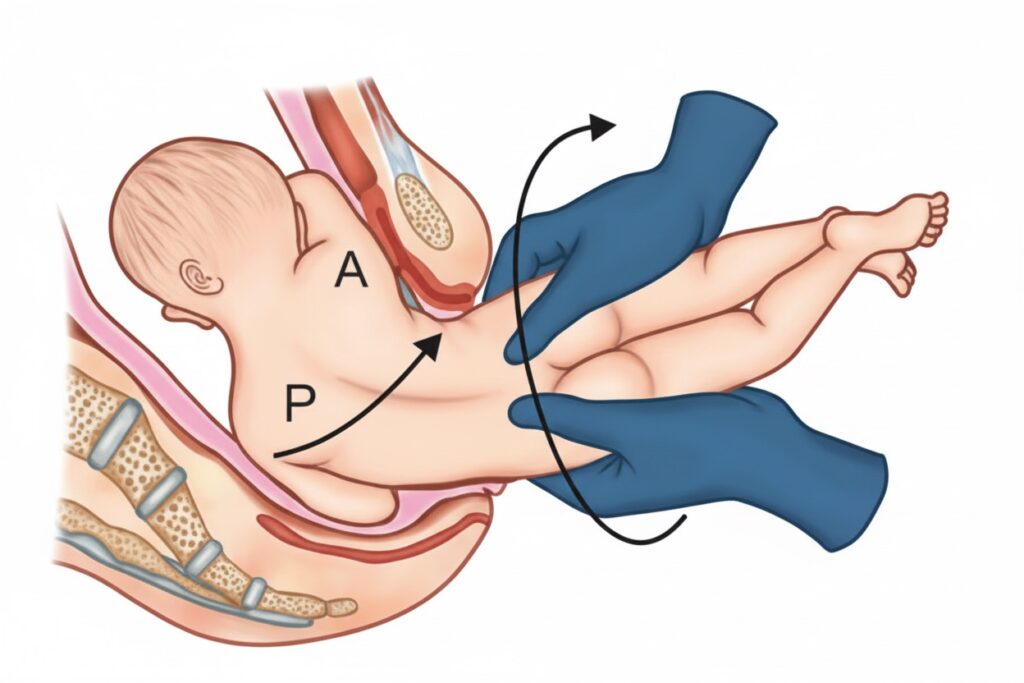
TCML

Apolipoprotein B48 and B100

  Feature Apolipoprotein B48 Apolipoprotein B100 Synthesissite Intestines (Enterocytes) Liver Length 48% shorter thanApolipoprotein B100 Full-length(4,536 amino acids) Amino acidSsequence Truncated form ofApolipoprotein B100 Full-length,longer sequence RNA Editing Result of RNA editing(cytidine to uridine conversion) No RNA editing,full-length protein Majorcomponent Chylomicron VLDL, IDL, LDL Function Involved in lipid transportfrom intestines Involved in lipidtransportfrom liver  

Apolipoprotein B48 and B100 Read More »

Trichomonas vaginalis

– Flagellated protozoan parasite  – Lack of cyst stage, it exists only in the trophozoite form – Symptoms: Profuse frothy greenish yellow discharge – It shows twitching motility / Jerky / Wobbling / Rotatory motility  🔴 NEET PG 2023– Colpitis macularis: Strawberry appearance of vaginal mucosa (Angry looking vagina) – Culture: Diamond’s media– Treatment: Metronidazole / Tinidazole CAMP

Trichomonas vaginalis Read More »

CAMP test and Reverse CAMP test

CAMP test-positive organism: 1. Streptococcus agalactiae (GBS/Group B Streptococcus)  2. Listeria monocytogenes CAMP test and Reverse CAMP test By TCML February 27, 2026 No Comments Structure locate between muscles of pharynx By TCML February 27, 2026 No Comments Oxygen-hemoglobin dissociation curve By TCML February 26, 2026 No Comments Third ventricle of brain By TCML February

CAMP test and Reverse CAMP test Read More »

Structure locate between muscles of pharynx

Anatomical gap Structures Passing Through Between Skull Base& SC – Eustachian tube – Levator veli palatini muscle – Ascending palatine artery – Palatine branch of ascending pharyngealartery  Between SC& MC – Stylopharyngeus muscle (supplied by CN-9) – Glossopharyngeal nerve (CN-9)  Between MC& IC (Thyrohyoidmembrane) – Internal laryngeal nerve (ILN) – Superior laryngeal artery & vein

Structure locate between muscles of pharynx Read More »

Epiploic foramen

– Aka Foramen of winslow – It’s an opening that connects the greater sac (general peritoneal cavity) with the lesser sac (omental bursa).  Boundaries Structure  Anterior Hepatoduodenal ligament, which contains portal triad- • Hepatic artery proper  • Portal vein  • Common bile duct (CBD) Posterior  • Inferior vena cava (IVC) • T12 vertebra  Superior  Caudate lobe of the liver  Inferior

Epiploic foramen Read More »

Pemphigus vulgaris

• Type: Autoimmune blistering disorder  • Autoantibodies: Against Desmoglein-3 (mucosa) ± Desmoglein-1 (skin) — components of desmosomes • Hypersensitivity type: Type 3 Hypersensitivity (Ag-Ab complex) • Blister type- Flaccid, Intraepidermal blisters    Pemphigus group of disorders  Pemphigoid group of disorders Bullae  Flaccid bullae Tense bullae (because whole epidermis  is present in bulla covering  so bulla is tight / tense) Bullae 

Pemphigus vulgaris Read More »

Oxygen-hemoglobin dissociation curve

  Shift Right Shift Left Shift Affection  for oxygen Decreased affinity Increased affinity Hb-oxygen  binding Oxygen is less tightly bound; more readily released Oxygen is more tightly bound;  less readily released  Causes – Increased CO2 – Decreased CO2   – Increased temperature – Decreased temperature   – Decreased pH (acidosis) – Increased pH (alkalosis)  

Oxygen-hemoglobin dissociation curve Read More »

Third ventricle of brain

• Location: between the right and left thalami. • Extends from the interventricular foramen (of Monro) anteriorly → to the cerebral aqueduct (of Sylvius)posteriorly. Boundaries Formed by Roof • Choroid plexus • Fornix Floor Formed by hypothalmic structure-  • Optic chiasma • Infundibulum  • Tuber cinereum • Mammillary bodies  • Posterior perforated substance • Tegmentum of midbrain  Lateral wall • Thalamus (superior part) • Hypothalamus (inferior part) Anterior wall Anterior commissure

Third ventricle of brain Read More »

Retinal detachment

Separation of RPE (Retinal pigmented epithelium) from neurosensory retina. Retinal detachment (RD) Description Examination finding Rhegmatogenous RD (Rhegma = Break) Retina continuity break Tear (due to traction)- • Horseshoe shaped RD • M/C cause is PVD  • PVD is physiological (normal) in old age Hole (after degeneration)-  • Lattice degeneration • Snail track degeneration • Corrugated appearance  • Schaffer’s sign  / Tobacco dust • Undulating

Retinal detachment Read More »