Topic Overview-
1. Boundaries
2. Content
3. Clinicals
4. Additional topics
Axilla is present between lateral part of chest wall and upper part of arm.
It is a pyramid shape structure that’s why we study axilla boundary into apex, base and four wall.

1. Boundaries-
Apex –
Apex of axilla is passage between neck and axilla that’s why it is called cervicoaxillary canal.
(See diagram below)
1. Anterior side – Clavicle (Posterior surface)
2. Posterior side – Scapula Bone (Superior border) and coronoid process
3. Medial side – First Rib (Outer border)

Base –
The part in which we apply deodorant (deo), that part is axilla base.
Axilla base boundaries-
1. Anterior side – Anterior axillary fold
2. Posterior side – Posterior axillary fold
3. Medial side – Chest wall
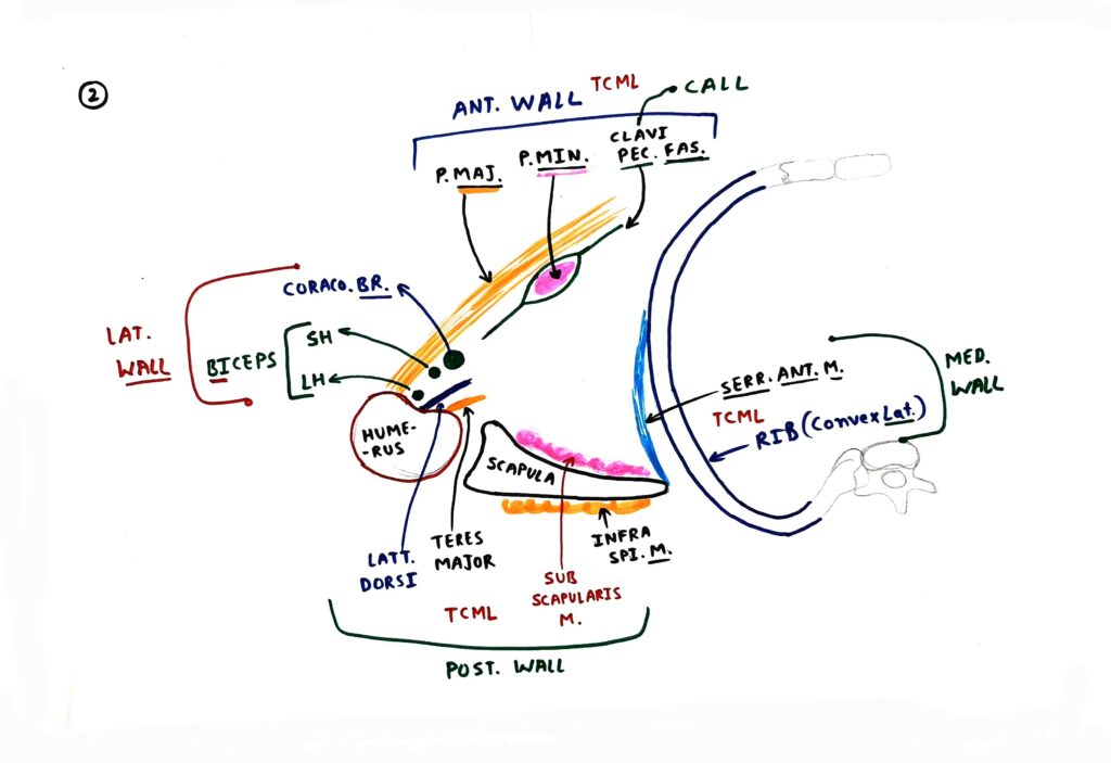
Four wall
Anterior wall-
Below structure are present on anterior side of axilla.
1. Pectoralis major muscle- Supply by medial and lateral pectoral nerve.
2. Pectoralis minor muscle – It is mainly supplied by medial pectoral nerve but some fibers of lateral pectoral nerve also supply it.
3. Clavipectoral fascia
Posterior wall-
1. Lattismus dorsi muscle – It is supply by thoracodorsal nerve
2. Teres major – It is supply by upper subscapular nerve.
3. Subscapularis muscle – It is supply by upper and lower subscapular nerve.
Lateral wall-
1. Biceps brachii muscle (Long and Short Head)
2. Coracobrachialis muscle
Both muscles are supplied by Musculocutaneous nerve.
Medial wall-
1. Serratus anterior – It is supplied by long thoracic nerve.
2. Ribs (Upper four ribs and their intercostal space)

2. Content-
1. Axillary artery
2. Axillary vein
3. Brachial Plexus cord
Lateral cord
1. Lateral pectoral nerve – Supply to pectoralis major and minor muscle.
Medial cord
1. Medial pectoral nerve – Supply to pectoralis major and minor muscle.
Posterior cord
1. Upper subscapular nerve – Supply to subscapularis and teres major muscle.
2. Lower subscapular nerve – Supply to subscapularis muscle.
4. Axillary lymph nodes
a. Anterior group
b. Posterior group
c. Lateral group
d. Central group
e. Apical group
5. Long thoracic nerve- It is supply to the serratus anterior muscle.
3. Clinicals-
1. Winging of scapula – It is due to damage of long thoracic nerve.
Long thoracic nerve supply to the serratus anterior muscle.
Serratus anterior muscle attached on medial border and inferior angle of scapula, so in the case of serratus anterior muscle palsy medial border and inferior angle of scapula is elevated.
NOTE :
Serratus anterior muscle is also called boxer’s muscle.
Long thoracic nerve is also called nerve of bell.
2. Breast cancer metastasis- Breast cancer is metastasis through axillary lymph nodes.
3. Breast Cancer Surgery (Mastectomy)-
Long thoracic nerve is injured in the breast cancer surgery.
Suspected lymph nodes are removed in breast cancer surgery.
4. Chest tube or intercostal drain –
Chest tube is used to drain pneumothorax, haemorhorax, pleural effusion etc.
It is insert in intercostal space.
Axillary lines are used to determine where to put the chest tube.
5. ECG Leads –
1. 5th ECG lead is placed in the anterior axillary line (5th intercostal space)
2. 6th ECG lead is placed in the middle axillary line (5th intercostal space)

4. Additional topics-
1. Clavipectoral fascia – Structure that pierce clavipectoral fascia
Mnemonic : CALL
C – Cephalic vein
A – Artery: Thoracoacromial artery
L – Lateral pectoral nerve
L – Lymphatics
2. Bicipital groove – Attachment and content
● Attachment-
Mnemonic : Lala between two major bodyguard
1. Lattismus dorsi muscle
2. Pectoralis major muscle
3. Teres major muscle
● Content-
1. Long head of Biceps brachii muscle
2. Ascending branch of ACHA (Anterior Circumflex Humeral Artery)
3. Brachial plexus cord –
(See axilla content above)
4. Axillary lymph nodes-
(See axilla content above)
5. Coracoid process – Attachment
1. Pectoralis minor muscle
2. Coracobrachialis muscle
3. Short head of biceps brachii muscle


