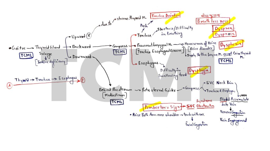
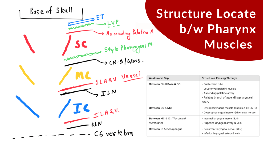
Head and neck anatomy

Tongue nerve supply

We study tongue nerve supply into two parts. 1. Sensory supply- 1. Anterior 2/3 of tongue- General sensation (GS) – Trigeminal nerve carry GS from anterior 2/3 of tongue. Special sensation (SS) – Facial nerve carry SS from anterior 2/3 of tongue. 2. Posterior 1/3 of tongue- Glossopharyngeal nerve carry both GS and SS from […]

Tongue nerve supply Read More »

SubarachnoId hemorrhage

Overview – 1. Subarachnoid space 2. Cause 3. Clinical features 4. Diagnosis ● Subarachnoid space – It is present between arachnoid and piamater. ● Circle of willis (COW) and CSF both are present in subarachnoid space (Inter peduncular cistern) NOTE – Inter peduncular cistern is a subarachnoid space and it contain circle of willis. ●

SubarachnoId hemorrhage Read More »

Thyroid gland

Overview- 1. Dyspnea 2. Dysphonia 3. Dysphagia 4. Tracheal Deviation 5. Retrosternal goiter 6. Pemberton’s Sign Thyroid gland present in neck region and it have two lobe (Right and left) and both lobe are connect with isthmus. Thyroid gland present in front of the tracha and it secret three hormone 1. Thyroxine or tetra iodo

Thyroid gland Read More »

Brain blood supply

Overview- 1. Vertebral Artery- 4th part (Five branches)2. Basilar Artery- Five branches 3. Internal Carotid Artery (ICA)- 4th part (Five branches)4. Circle of Willis (COW)- Draw hexagon 5. Cerebellum blood supply- Three artery 6. Clinicals- Stroke, Ataxia, PICA syndrome (Wallenberg syndrome or Lateral medullary syndrome) NOTE: Vertebral, and Internal carotid artery- • Both artery are

Brain blood supply Read More »

Pterion

Overview- We covered pterion anatomy into below points. 1. Fontanelle- Pterion2. Location- Temporal fossa3. Formation 4. Artery- Middle meningeal artery 5. Nerve- Auriculotemporal nerve6. Hematoma7. Surgical landmark 8. Fracture 1. In pediatrics we read four fontanelle- A. Anterior fontanelle or BregmaB. Posterior fontanelle or LambdaC. Anterolateral fontanelle or Pterion D. Posterolateral fontanelle or Asterion Mnemonic:

Pterion Read More »

Temporal fossa

Overview- 1. Boundaries- Anterior, posterior, superior, inferior, lateral, and medial. 2. Communication- Infratemporal fossa. 3. Content- Muscle, artery, and nerve. ● It is a fan shape, shallow fossa. ● Two in number and present on anterolateral side of the skull. ● Temporalis muscle (Fan shape muscle) are attached on it. 1. Boundaries- The boundaries of

Temporal fossa Read More »

Larynx Anatomy

Overview- In this article we covered- • Larynx Intrinsic muscles (Total 9 muscles) • Vagus nerve (Superior and inferior/recurrent laryngeal nerve) • Pharyngeal arch (4th and 6th arch) Larynx muscles (Intrinsic muscles) ● Development– All Intrinsic Muscles of the Larynx are developed from the 6th pharyngeal arch except the Cricothyroid muscle (this is developed from

Larynx Anatomy Read More »

Carotid artery

OVERVIEW- In this article we covered- A. External carotid artery and its branchesB. Maxillary artery and its branches C. Maxillary nerve (Trigeminal nerve) and its branches Branches of External carotid artery (ECA) • this is the branch of the common carotid artery (Common carotid artery divide into two part; external and internal carotid artery- see

Carotid artery Read More »