Overview –
1. Theory
2. Diagram
3. Clinical
This is a aponeurotic sheath covering the rectus abdominis muscle (Anterior abdominal wall muscle). We covered rectus sheath anatomy into three point are as follow: Theory, diagram and clinical’s.
A. Theory –
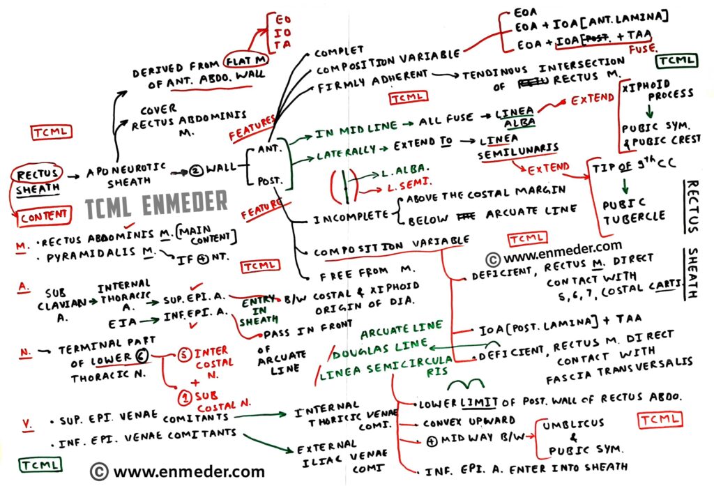
The rectus sheath has two wall, and it contain two muscles, two artery, two vein, and six nerve.
1. Wall of rectus sheath-
Rectus sheath has two wall anterior and posterior. (Fig. Transverse section)
NOTE: Both wall of rectus sheath has different composition. (for more detail see transverse section of rectus sheath that are given below in this blog)
2. Content-
(a) Two muscle:
Rectus abdominis muscle (This is main content of rectus sheath) and pyramidalis muscle (If present).
(b) Two artery (Fig. Sagittal section):
Superior epigastric artery (This is the branch of internal thoracic artery) and inferior epigastric artery (This is the branch of external iliac artery).
(Abdominal aorta– common iliac artery- external iliac artery- inferior epigastric artery)
(c) Two vein:
Superior epigastric vein (Drain into internal thoracic vein) and inferior epigastric vein (Drain into external epigastric vein).
(d) Six nerve:
Lower six thoracic nerve (Five intercostal nerve and sub costal nerve)

B. Diagram-
To understand rectus sheath anatomy, we cut the rectus sheath into below two section. (Figs. Sagittal and transverse section)
1. Sagittal section:

Through this we easily understand anterior and posterior wall of rectus sheath and which structure are present on anterior and posterior side.
2. Transverse section (T.S.):
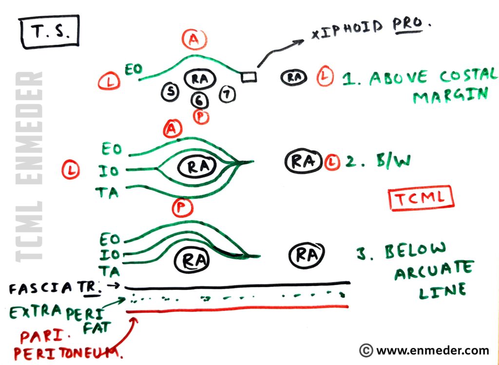
Here we draw three tranverse section diagram of rectus sheath to understand the composition of rectus sheath wall.
(a) Above the costal margin (Fig. Transverse section):
● Anterior wall:
It is formed by external oblique aponeurosis (EOA).
● Posterior wall:
In this part (above costal margin) posterior wall is absent and the rectus abdominis muscle are present just anterior to the 5th, 6th, and 7th costal cartilage.
(Aponeurosis are flat structure and tendon are cord like structure)
(b) Between costal margin and arcuate line (Fig. Transverse section):
● Anterior wall:
It is formed by external oblique aponeurosis and internal oblique aponeurosis. (Anterior lamina)
● Posterior wall:
It is formed by internal oblique aponeurosis (Posterior lamina) and transversus abdominis aponeurosis.
(c) Below the arcuate line (Fig. Transverse section):
● Anterior wall:
It is formed by external, internal oblique and transversus abdominis aponeurosis.
(NOTE: External and Internal abdominis aponeurosis are fused and transverse abdominis aponeurosis remains separate)
● Posterior wall:
In this part (below arcuate line) posterior wall is absent and the rectus abdominis muscle are present just anterior to the fascia tranversalis.

C. CLINICAL’S-
1. Recti divarication
2. Rectus sheath hematoma
3. Hernia
📌 All TCML Hand written notes/charts and videos are available on TCML Mobile App. (FREE for All Users)
🔴 To download click on below pic.


