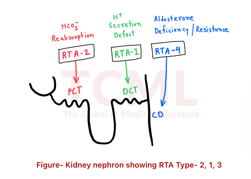
Renal tubular acidosis
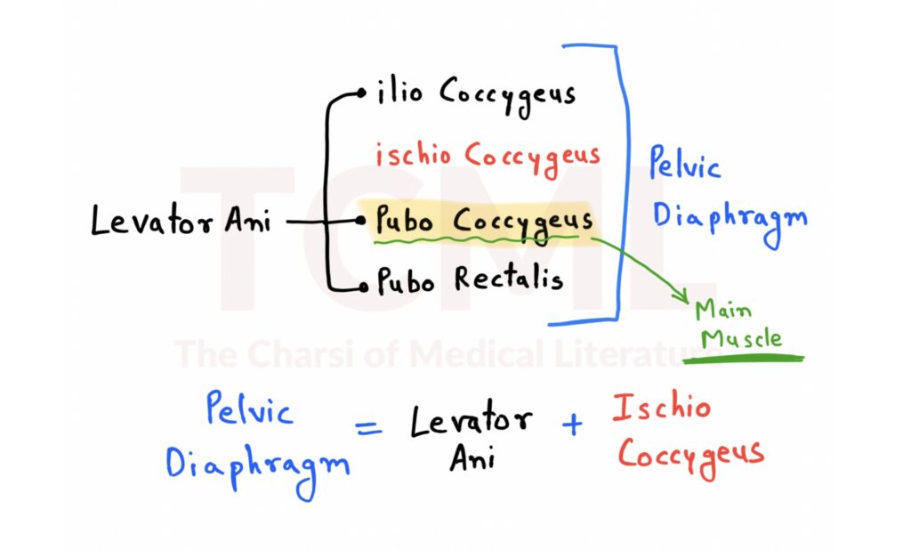
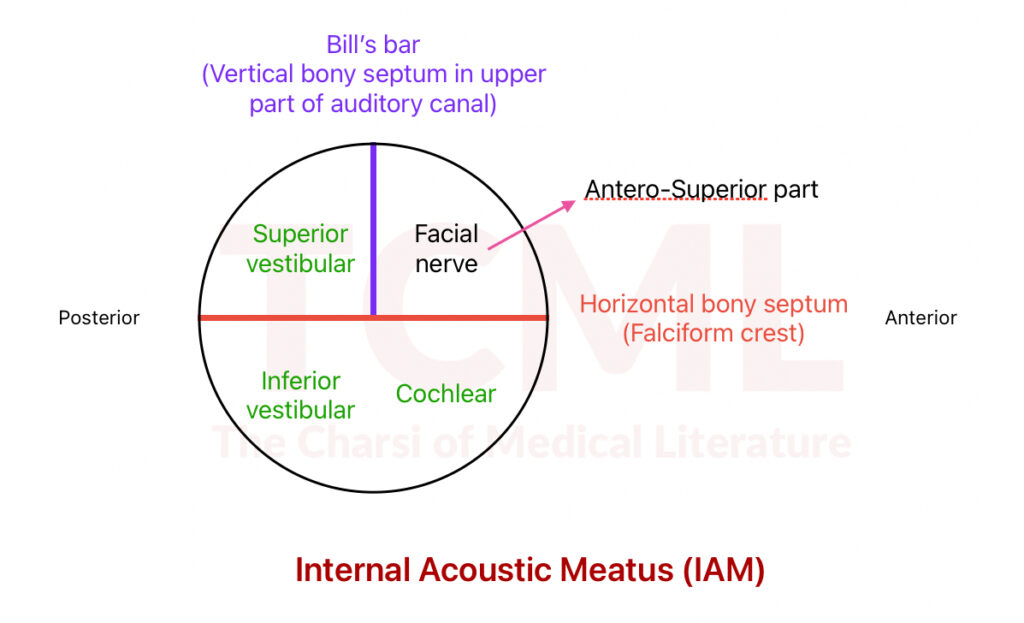
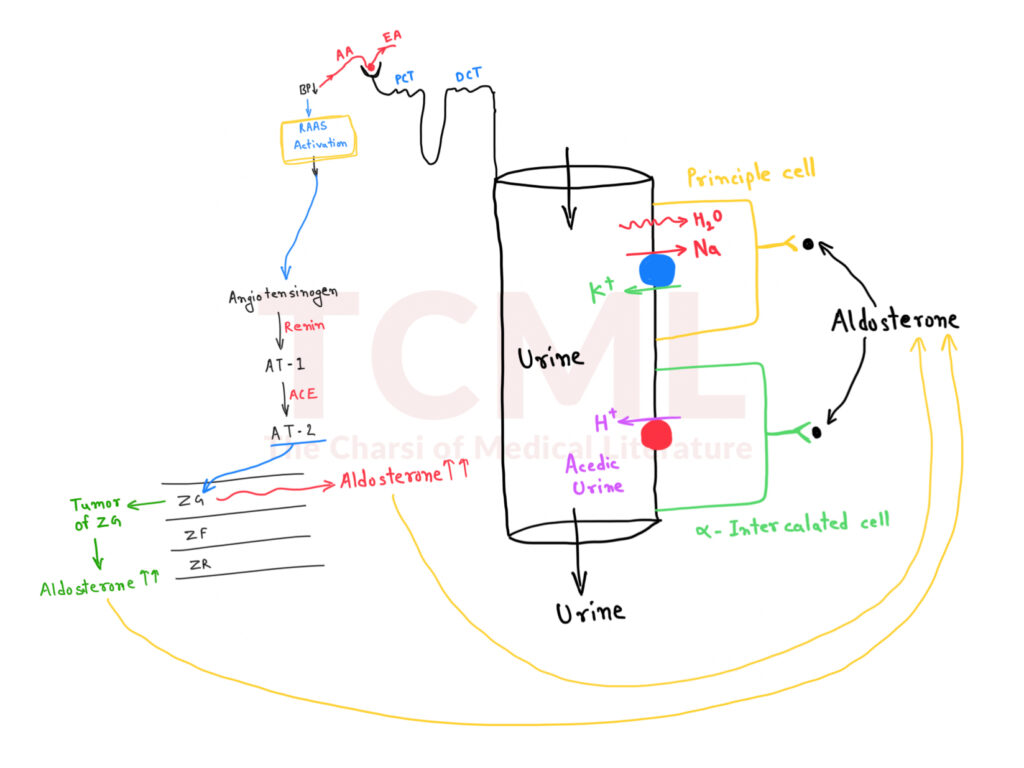
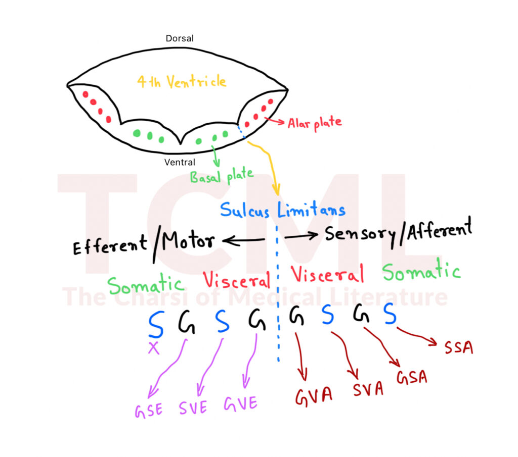
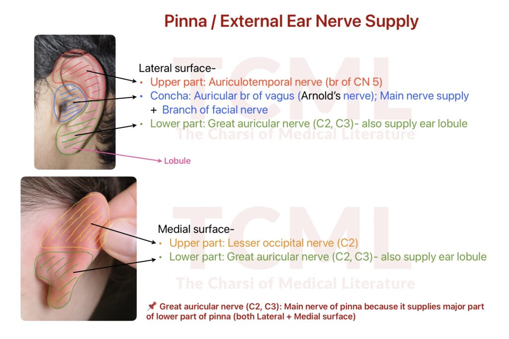
Pelvic diaphragm By TCML February 20, 2026 No Comments Internal acoustic meatus By TCML February 20, 2026 No Comments Hyperaldosteronism, Conn syndrome By TCML February 20, 2026 No Comments Functional column of cranial nerve By TCML February 20, 2026 No Comments External ear nerve supply By TCML February 20, 2026 No Comments
Renal tubular acidosis Read More »