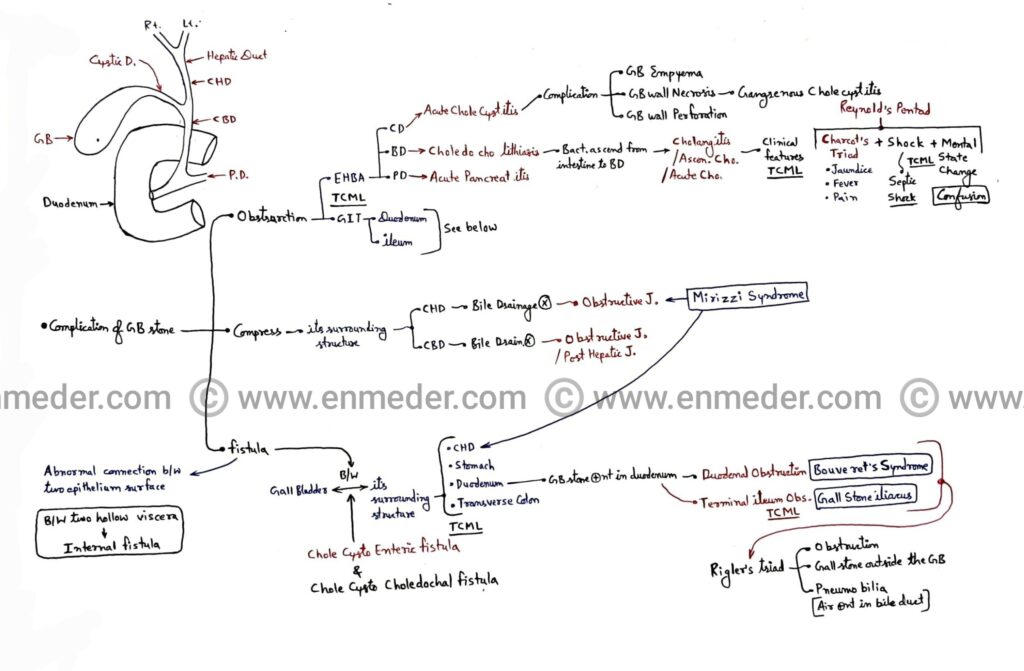
neet pg

Anatomy questions

Overview- 1. Blood Supply 2. Anastomosis 3. Venous drainage 4. Covering 5. Nerve and plexus 6. Retinaculum 7. Boundaries and content 8. Joints 9. Viscera 10. NeuroAnatomy 11. Embryology 12. Histology 1. Blood Supply 1. Aorta parts and it’s branches 2. Heart blood supply (Aortic sinus) – Right and left coronary artery Question. Myocardial infraction […]

Anatomy questions Read More »

GI bleeding

Overview- 1. Epistaxis 2. Hemoptysis 3. Hematemesis 4. Hematochezia 5. Melena6. Hematouria Blood from Nose (Epistaxis) It is also called Epistaxis. Some important questions about nose bleeding. Question 1. Epistaxis is due to the rupture of Kiesselbach’s plexus which is present at little’s area of anterior inferior part of the nasal septum. Question 2. Four

GI bleeding Read More »

Thyroid gland

Overview- 1. Dyspnea 2. Dysphonia 3. Dysphagia 4. Tracheal Deviation 5. Retrosternal goiter 6. Pemberton’s Sign Thyroid gland present in neck region and it have two lobe (Right and left) and both lobe are connect with isthmus. Thyroid gland present in front of the tracha and it secret three hormone 1. Thyroxine or tetra iodo

Thyroid gland Read More »

Brain blood supply

Overview- 1. Vertebral Artery- 4th part (Five branches)2. Basilar Artery- Five branches 3. Internal Carotid Artery (ICA)- 4th part (Five branches)4. Circle of Willis (COW)- Draw hexagon 5. Cerebellum blood supply- Three artery 6. Clinicals- Stroke, Ataxia, PICA syndrome (Wallenberg syndrome or Lateral medullary syndrome) NOTE: Vertebral, and Internal carotid artery- • Both artery are

Brain blood supply Read More »

Pterion

Overview- We covered pterion anatomy into below points. 1. Fontanelle- Pterion2. Location- Temporal fossa3. Formation 4. Artery- Middle meningeal artery 5. Nerve- Auriculotemporal nerve6. Hematoma7. Surgical landmark 8. Fracture 1. In pediatrics we read four fontanelle- A. Anterior fontanelle or BregmaB. Posterior fontanelle or LambdaC. Anterolateral fontanelle or Pterion D. Posterolateral fontanelle or Asterion Mnemonic:

Pterion Read More »

Temporal fossa

Overview- 1. Boundaries- Anterior, posterior, superior, inferior, lateral, and medial. 2. Communication- Infratemporal fossa. 3. Content- Muscle, artery, and nerve. ● It is a fan shape, shallow fossa. ● Two in number and present on anterolateral side of the skull. ● Temporalis muscle (Fan shape muscle) are attached on it. 1. Boundaries- The boundaries of

Temporal fossa Read More »

Cubital Fossa

Overview 1. Features- Shape, Location & Homologous 2. Boundaries- Lateral, Medial, Apex, Floor, Roof & Floor3. Content- M B T R4. Clinicals- Blood Pressure Measure, Brachial Pulse, Intra Venous Injection, Collection of Blood Sample, Fracture, Volkmann’s Ischemic Contracture, 1. FEATURES This is triangular in shape- So it has three border- Lateral, Medial, Lower ( Base

Cubital Fossa Read More »

Bell’s Palsy

Overview:  In this topic we covered – Facial Nerve branches Bell’s Palsy Bell’s Phenomenon Lagophthalmos Exposure Keratopathy Corneal Ulcers Epiphora or Watering Eye Paralytic Ectropion Treatment Facial Nerve: Branches Overview Greater Petrosal Nerve: Join Deep Petrosal Nerve ( from Sympathetic nerve plexus around Internal Carotid Artery ) and form Nerve of Pterygoid Canal Br. to

Bell’s Palsy Read More »