
Scapular anastomosis
Anastomosis around scapula- 1. Anastomosis over acromion process 2. Anastomosis around body of scapula
Scapular anastomosis Read More »
![]()
![]()
Anastomosis around scapula- 1. Anastomosis over acromion process 2. Anastomosis around body of scapula
Scapular anastomosis Read More »
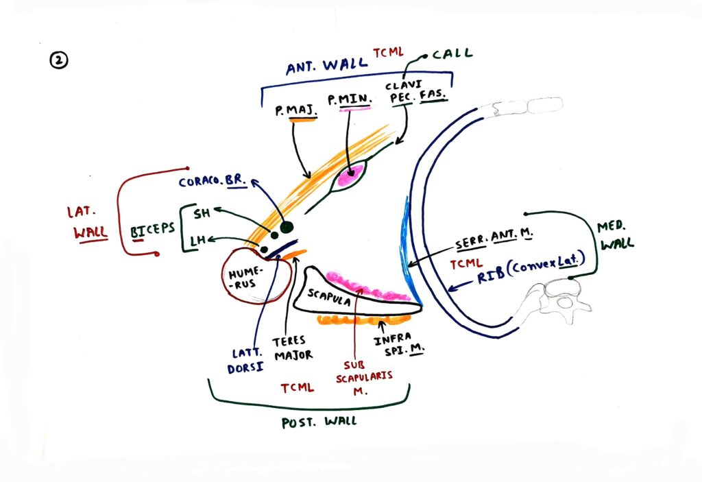
Topic Overview- 1. Boundaries 2. Content 3. Clinicals 4. Additional topics Axilla is present between lateral part of chest wall and upper part of arm. It is a pyramid shape structure that’s why we study axilla boundary into apex, base and four wall. 1. Boundaries- Apex –Apex of axilla is passage between neck and axilla that’s
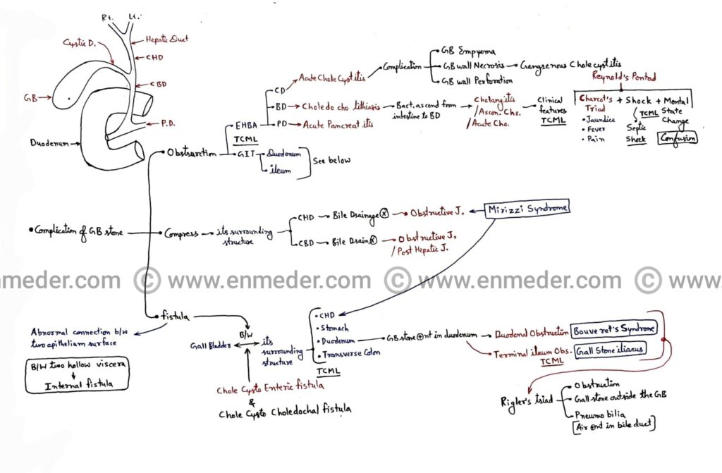
Complications of gallstone/cholelithiasis- 1. Obstruction of extra hepatic biliary apparatus and gastrointestinal tract 2. Compression of common hepatic duct and common bile duct 3. Fistula between gallbladder and it’s surrounding structures 1. Obstruction – A. Extra hepatic biliary apparatus (EHBA) – 1. Cystic duct (CD)Due to obstruction of cystic duct by gallstone it causes acute

Overview 1. Femoral nerve and it’s branches 2. Cutaneous nerve of thigh 3. Patellar plexus 4. Muscles of anterior compartment of thigh 5. Hip joint nerve supply 6. Knee joint nerve supply 7. Clinicals In lower limb anatomy we study five main nerves 1. Femoral nerve 2. Obturator nerve 3. Sciatic nerve 4. Tibial nerve

Overview- 1. Boundaries- Anterior, posterior, superior, inferior, lateral, and medial. 2. Communication- Infratemporal fossa. 3. Content- Muscle, artery, and nerve. ● It is a fan shape, shallow fossa. ● Two in number and present on anterolateral side of the skull. ● Temporalis muscle (Fan shape muscle) are attached on it. 1. Boundaries- The boundaries of
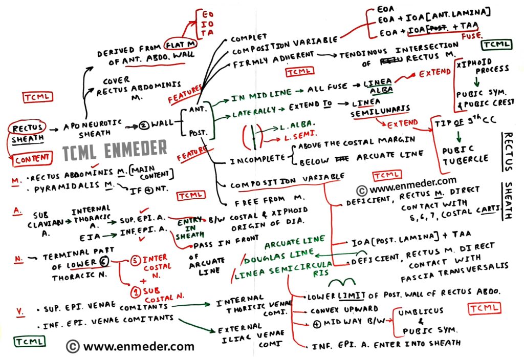
Overview – 1. Theory 2. Diagram 3. Clinical This is a aponeurotic sheath covering the rectus abdominis muscle (Anterior abdominal wall muscle). We covered rectus sheath anatomy into three point are as follow: Theory, diagram and clinical’s. A. Theory – The rectus sheath has two wall, and it contain two muscles, two artery, two vein,

Hello future Dr. I am Pawan nagar from TCML Team. A lot of medical students messaged me (On the YouTube comment section, WhatsApp, Instagram and Messenger etc.) and said that sir we do not understand anything in anatomy and we’re scared of anatomy. Based on your messages we have created a sequence of six charts

Overview- In this article we covered. • Quadrangular Space • Upper Triangular Space • Lower Triangular Space • Axillary Nerve • Lower Subscapular Nerve • Radial Nerve • Axillary Artery branches (PCHA and Circumflex scapular artery) • Brachial Artery branches (Profunda or Deep brachii artery) Mnemonic- 3 Spaces, 3 Muscles, and 3 Artery. 3 SPACES:
Intermuscular Spaces Read More »

Overview- In this article we covered- • Larynx Intrinsic muscles (Total 9 muscles) • Vagus nerve (Superior and inferior/recurrent laryngeal nerve) • Pharyngeal arch (4th and 6th arch) Larynx muscles (Intrinsic muscles) ● Development– All Intrinsic Muscles of the Larynx are developed from the 6th pharyngeal arch except the Cricothyroid muscle (this is developed from

OVERVIEW- In this article we covered- A. External carotid artery and its branchesB. Maxillary artery and its branches C. Maxillary nerve (Trigeminal nerve) and its branches Branches of External carotid artery (ECA) • this is the branch of the common carotid artery (Common carotid artery divide into two part; external and internal carotid artery- see