
Eyeball
Eyeball A. Coats of eyeball – 1. Fibrous coat 2. Vascular coat 3. Nervous coat B. Segments and Chambers – 1. Anterior segment – It is divide into two chambers. Anterior chamber Posterior chamber 2. Posterior segment –
![]()
![]()
Eyeball A. Coats of eyeball – 1. Fibrous coat 2. Vascular coat 3. Nervous coat B. Segments and Chambers – 1. Anterior segment – It is divide into two chambers. Anterior chamber Posterior chamber 2. Posterior segment –
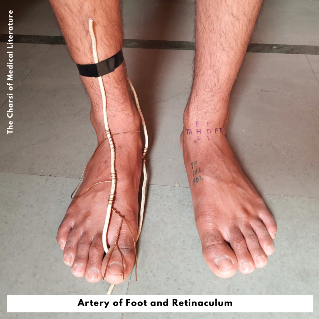
Dorsalis pedis artery branches- 1. Tarsal artery – Lateral and medial 2. Arcuate artery of foot 3. 1st dorsal metatarsal artery4. Deep plantar artery
Dorsalis pedis artery Read More »

There are 12 pair of cranial nerve are present in our body. 1. Ophthalmic nerve 2. Optic nerve 3. Oculomotor nerve – It supplies to all eye muscles except superior oblique and lateral rectus. 4. Trochlear nerve – It supplies only one eye muscle, superior oblique muscle (SO)5. Trigeminal nerve 6. Abducens nerve – It

Chart Highlights- 1. Femoral triangle 2. Femoral sheath – It is divided into three compartment (Lateral, intermediate and lateral)3. Femoral canal – It is the medial compartment of femoral sheath.4. Femoral ring – It is the base of femoral canal.
Femoral triangle, sheath and canal Read More »

Chart Highlights- 1. Shoulder joint relation 2. Coraco acromial arch 3. Musculo tendinious cuff 4. Bicipital groove 5. Axillary artery (1st rib, pectoralis minor, and teres major)

Chart Highlights- 1. Coronoid process 2. Coronoid fossa 3. Coranoid process 4. Olecranon process 5. Olecranon fossa 6. Trochlear notch 7. Trochlea 8. Radial notch 9. Radial fossa


